Humerus fractures (upper extremity)
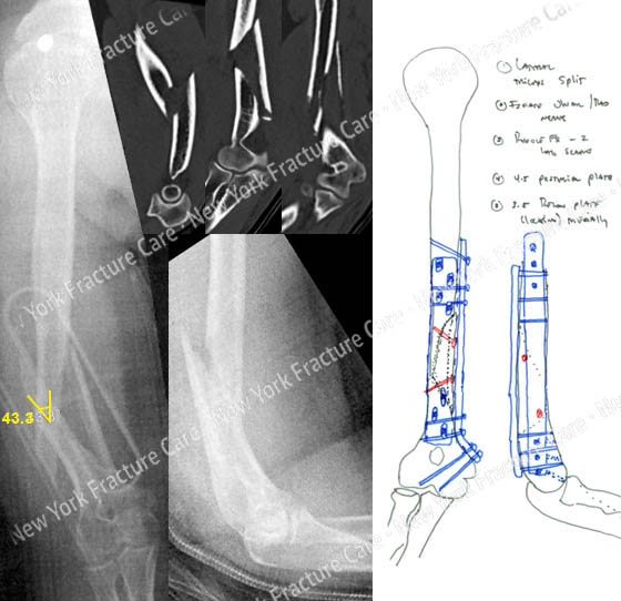
A 58-year-old female slipped and fell and sustained a closed right-sided displaced humerus fracture with a medial butterfly fragment.
She was taken to an outside hospital and placed in a splint for initial management. A CT scan was also performed at the outside hospital. She was then referred to David L. Helfet, MD at the Orthopaedic Trauma Service of the Hospital for Special Surgery for definitive management.
Open reduction and internal fixation was planned and performed with placement of locking and reconstruction plates and screws placed in a 90/90 fashion.
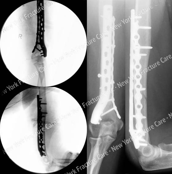
She returned for follow-up visits and radiographs at 6 months following surgery revealed a healed humerus fracture in excellent alignment with maintenance of reduction and fixation and she has returned to pre-injury activities and reports resolution of pain.
-
Figure A
Anteroposterior (AP) and lateral radiographs reveal a displaced humeral fracture with a medial butterfly fragment and CT images further delineate the fracture pattern (top images). Plate fixation in a 90/90 fashion was planned. -
Figure B
Intraoperative fluoroscopic AP and lateral images reveal adequate reduction and alignment (left images) and radiographs at 6 months reveal a healed humerus fracture in excellent alignment with maintenance of reduction and fixation (right images).
Tags: David Helfet MD, Humerus Fracture


