Complex fractures
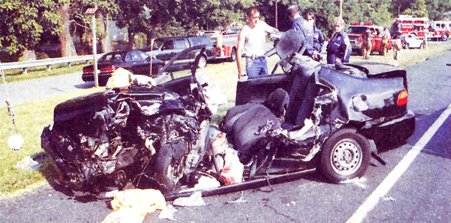
A 17-year-old male driver of an automobile was involved in a head-on collision with a tractor trailer. After being trapped inside the vehicle for approximately one hour, he was extricated and flown to a local trauma center along with the other passengers who sustained injuries of a lesser severity.
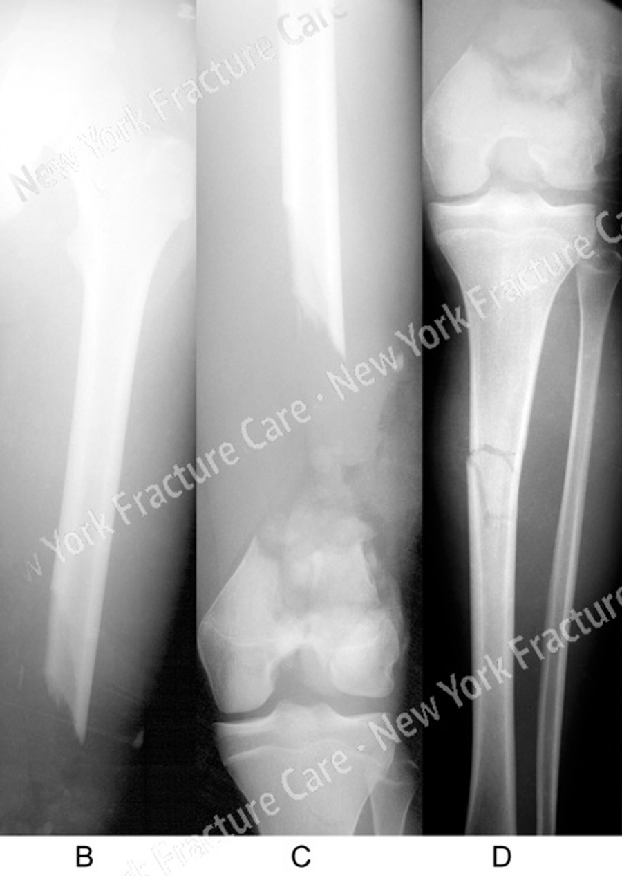
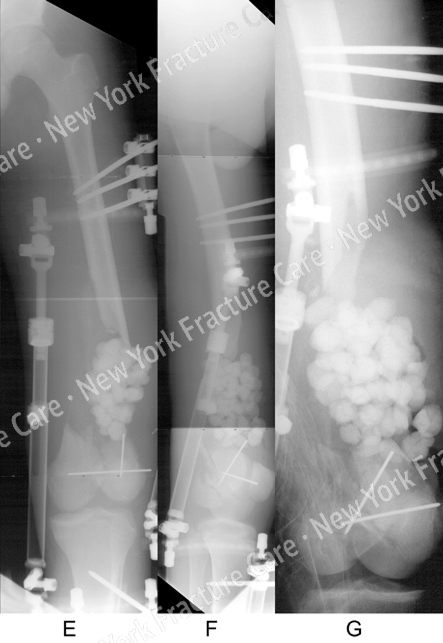
He was listed in critical condition and diagnosed with an open left-sided AO/OTA Type C3.3 distal femur fracture with segmental bone defect and an ipsilateral tibial shaft fracture. Spanning external fixation was placed for initial stabilization and antibiotic beads were subsequently placed in the femoral defect at 3 days following the injury. He was transferred to us for definitive management at 2 weeks after the injury.
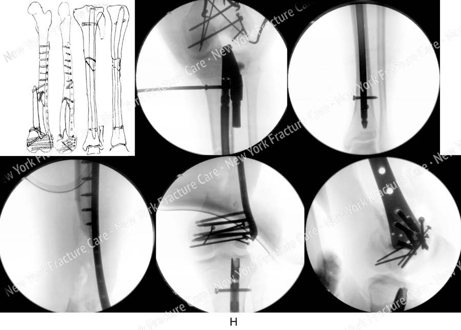
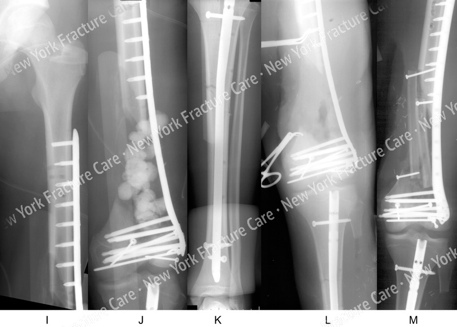
Open reduction and internal fixation (ORIF) was performed with placement of an intramedullary (IM) nail for treatment of the tibial shaft fracture and locking screws and then ORIF of the distal femur fracture was performed with placement of a Less Invasive Stabilization System (LISS) locking plate and screws.
One week later a second surgery was performed. The antibiotic beads were removed and the defect was prepared for bone graft placement. A second incision was made along the lateral border of the ipsilateral fibula and a free vascularized fibula bone graft was harvested for transplant to the femoral defect. It was docked in a double barrel fashion and stabilized using screw fixation.
Following surgery he returned for regular follow up visits. Three months after surgery all of the fractures were healing with incorporation of bone graft. The LISS plate was removed 4.5 years following the initial surgery.
The clinical and radiographic results were excellent including bony union, full range of motion and complete resolution of pain and return to pre-injury activities.
-
-
Figure B, C, D
Anteroposterior (AP) x-rays illustrating an AO/OTA Type C3.3 distal femur fracture with segmental bone defect and an ipsilateral tibial shaft fracture. -
Figure E, F, G
AP and lateral radiographs following placement of external fixation and antiobiotic beads at the site of the segmental bone defect. -
Figure H
(Counterclockwise from top-left) Preoperative plan, fluoroscopic images showing placement of intramedullary nail (IM) for the tibial shaft fracture and locking screws and open reduction and internal fixation (ORIF) of the distal femur fracture with placement of a LISS locking plate and screws. -
Figure I, J, K, L, M
Immediate postoperative radiographs demonstrating adequate fixation and alignment -
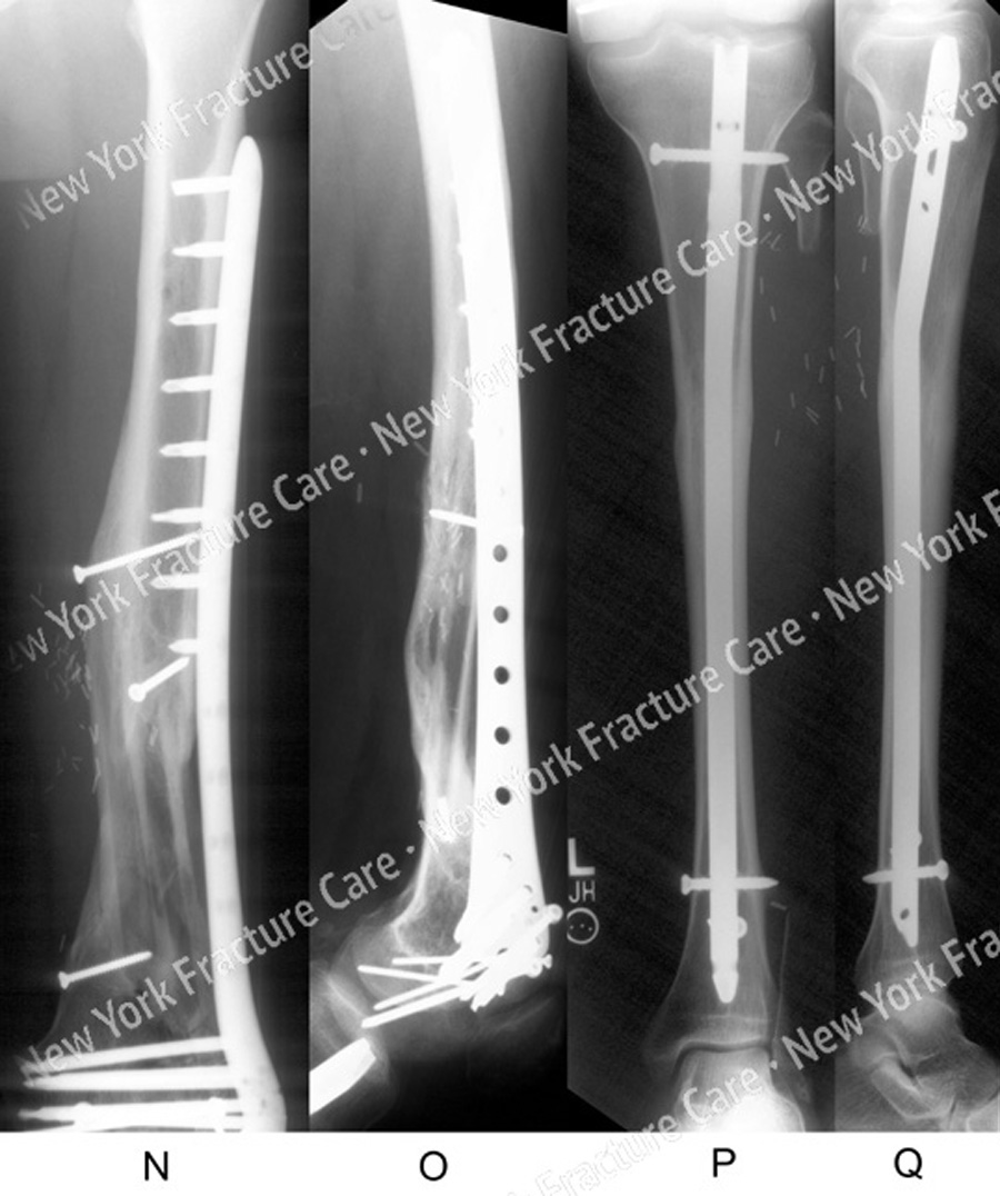
Figure N, O, P, Q
AP and lateral x-rays 3.5 years following ORIF showing healed a distal femur fracture with incorporation of the fibular bone graft and a healed tibial shaft fracture. -
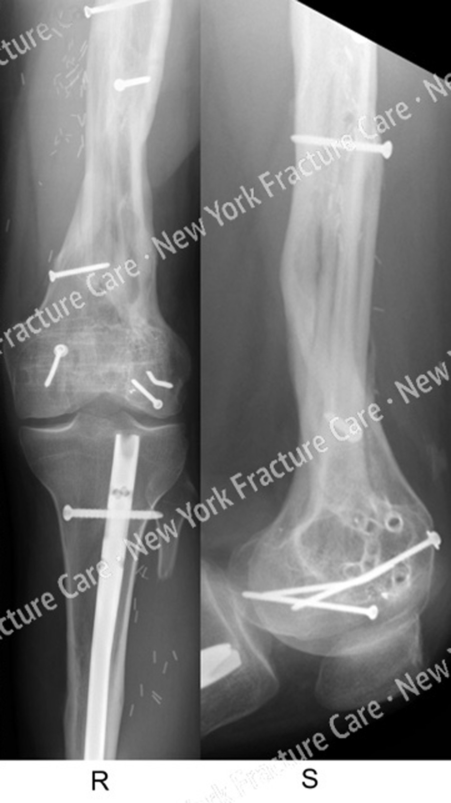
Figure R, S
AP and lateral x-rays 8 months following removal of LISS plate and screws and 4.5 years following fracture surgery.
Tags: Broken Femur, Tibia Fracture

