Femoral head fractures (lower extremity)
A 64-year-old female was involved as a passenger in a high-speed motor vehicle accident while on vacation in Nicaragua.
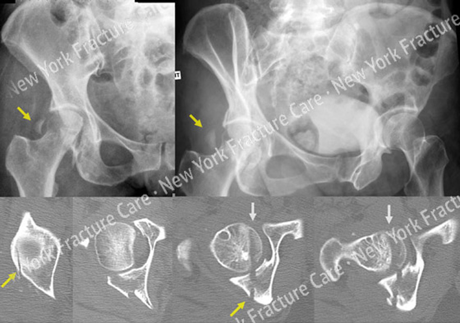
She was taken to a local hospital and diagnosed with a right-sided posterior hip dislocation that was closed reduced. She also had an associated right-sided Pipkin IV Type fracture of the femoral head with associated Posterior Wall Type fracture of the acetabulum.
After placement of skeletal traction she was transferred to us through a medical flight facilitated through her travel insurance. Open reduction and internal fixation (ORIF) was performed through a Kocher-Langenbeck surgical approach with an anterior surgical hip dislocation.
The Posterior Wall fracture was reduced and fixed with one-third tubular and pelvic reconstruction plates and screws. The femoral head fracture was reduced and fixed with sub-articular screws and preservation of the blood supply, reduction, hip stability, and adequate hardware placement was confirmed.
At the time of her latest follow-up at 5 months radiographs reveal healed acetabular and femoral head fractures and maintenance of reduction with mild degenerative changes in the hip joint.
-
Figure A
X-Rays (top) revealing a right-sided Pipkin IV femoral head fracture and associated Posterior Wall acetabular fracture (yellow arrows) and CT-Scan images (bottom) further delineating the fracture patterns (femoral head fracture is indicated with grey arrows). -
Figure B
Preoperative plan (left) and post-operative CT-Scan images (right) 1 day following ORIF. -
Figure C
Radiographs at 5-months post-operative revealing healed femoral head and acetabular fractures.
Tags: Hip Dislocation


