Fracture treatment in older patients
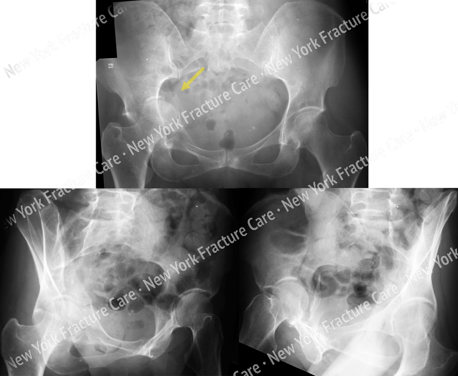
A 63-year-old female fell from a standing height. Radiographs taken at the outside hospital revealed an Anterior Column Type acetabular fracture with displacement of the quadrilateral surface and medial femoral head protrusion.
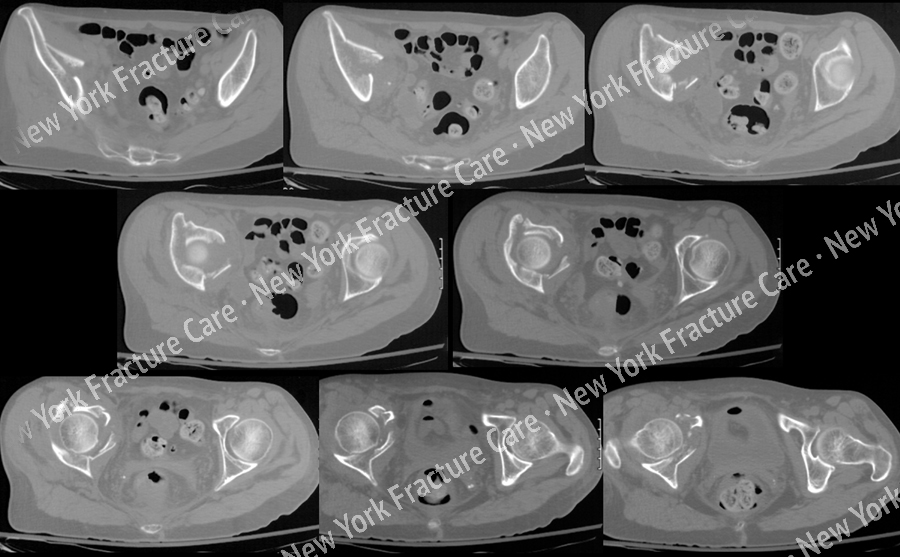
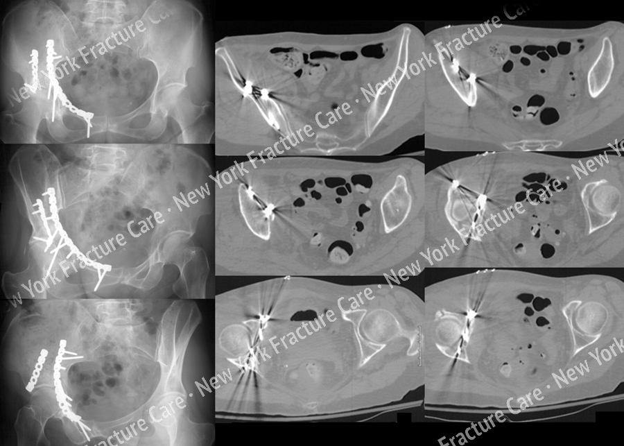
A CT-Scan was also performed to further delineate the fracture pattern. She was transferred to the us for definitive management. Open reduction and internal fixation (ORIF) was performed through an ilioinguinal approach with placement of plates and screws.
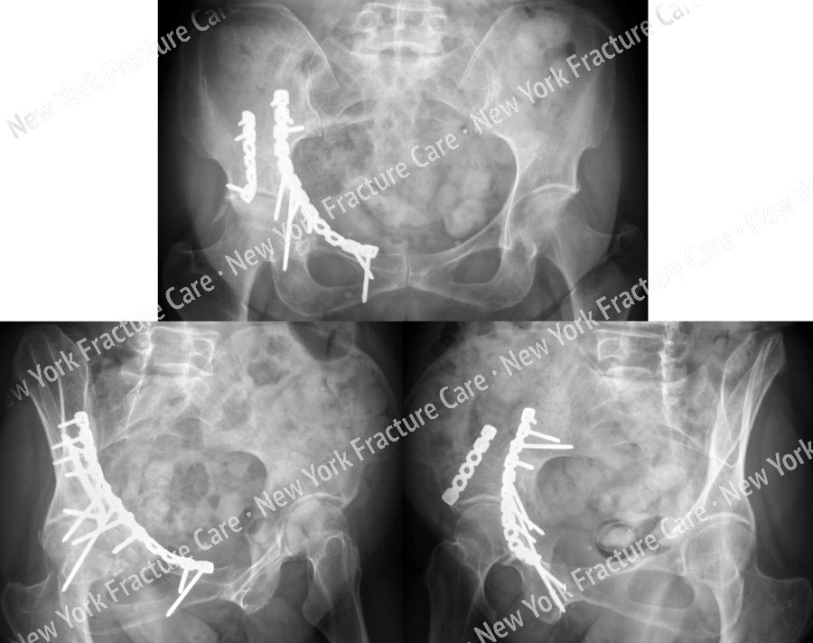
The patient continued to return for regular follow-up visits and had an excellent result. At her most recent follow-up visit at 9 years she has excellent clinical and radiographic results and a full return to her pre-injury status and activities of daily living.
-
Figure A
Radiographs demonstrating a right-sided Anterior Column acetabular fracture with displacement of the quadrilateral surface and medial femoral head protrusion (arrow). -
-
Figure C
Postperative AP and Judet radiographs (left) and postoperative CT scan images illustrating a satisfactory reduction and placement of hardware. -
Figure D
Radiographs 9 years following surgery demonstrating an excellent result with maintenance of reduction.
Tags: Broken Femur


