Minimally invasive fracture surgery
A 34-year-old male fell while snowboarding and landed on his outstretched left upper extremity.
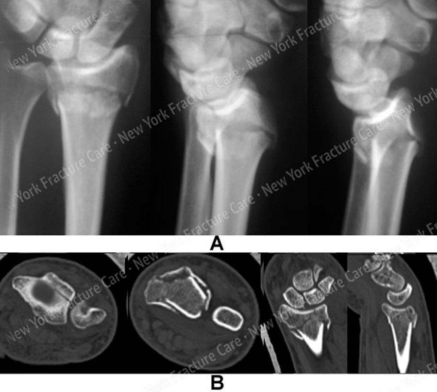
Radiographs at a local hospital revealed a left-sided unstable distal radius fracture with articular extension and dorsal comminution. A fracture splint was placed and he was referred to us for definitive management.
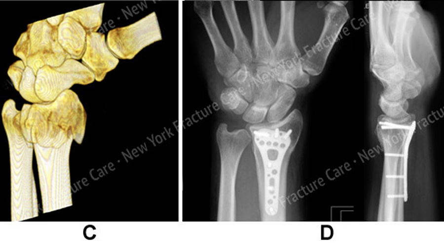
Open Reduction and Internal Fixation (ORIF) was performed with placement of a volar locking plate and screws and indirect reduction of the dorsal comminution.
He returned for regular follow-up and healed uneventfully, and at 6 months he presented with excellent results including a healed distal radius fracture in excellent alignment, full range of motion and resolution of pain and returned to all pre-injury activities and sports.
-
Figure A, B
(A) Anteroposterior (AP) and lateral injury radiographs revealing an unstable left-sided distal radius fracture with art icular extension and dorsal comminution, loss of volar tilt and radial inclination and height.
(B) CT scan further delineating fracture pattern.
-
Figure C, D
(C) 3D CT reconstruction scan image.
(D) Postoperative AP and lateral radiographs at 6 months illustrating a healed distal radius fracture in excellent alignment.
Tags: Broken Arm


