Stress fractures
A 20-year-old competitive track and field athlete at the collegiate level presented to us following an 18-month history of insidious onset of right-sided shin pain.
Her symptoms were aggravated by activity, especially the long jump. One year earlier she had been diagnosed with a stress fracture of her tibia and treated with a bone stimulator and physiotherapy modalities, including ultrasound. She had also tried a short period of immobilization in a CAM walker boot.
Following this treatment she had only temporary improvement in her symptoms and was referred to us. At the time of presentation she was experiencing significant right shin pain that was preventing her from competing in track and field events.
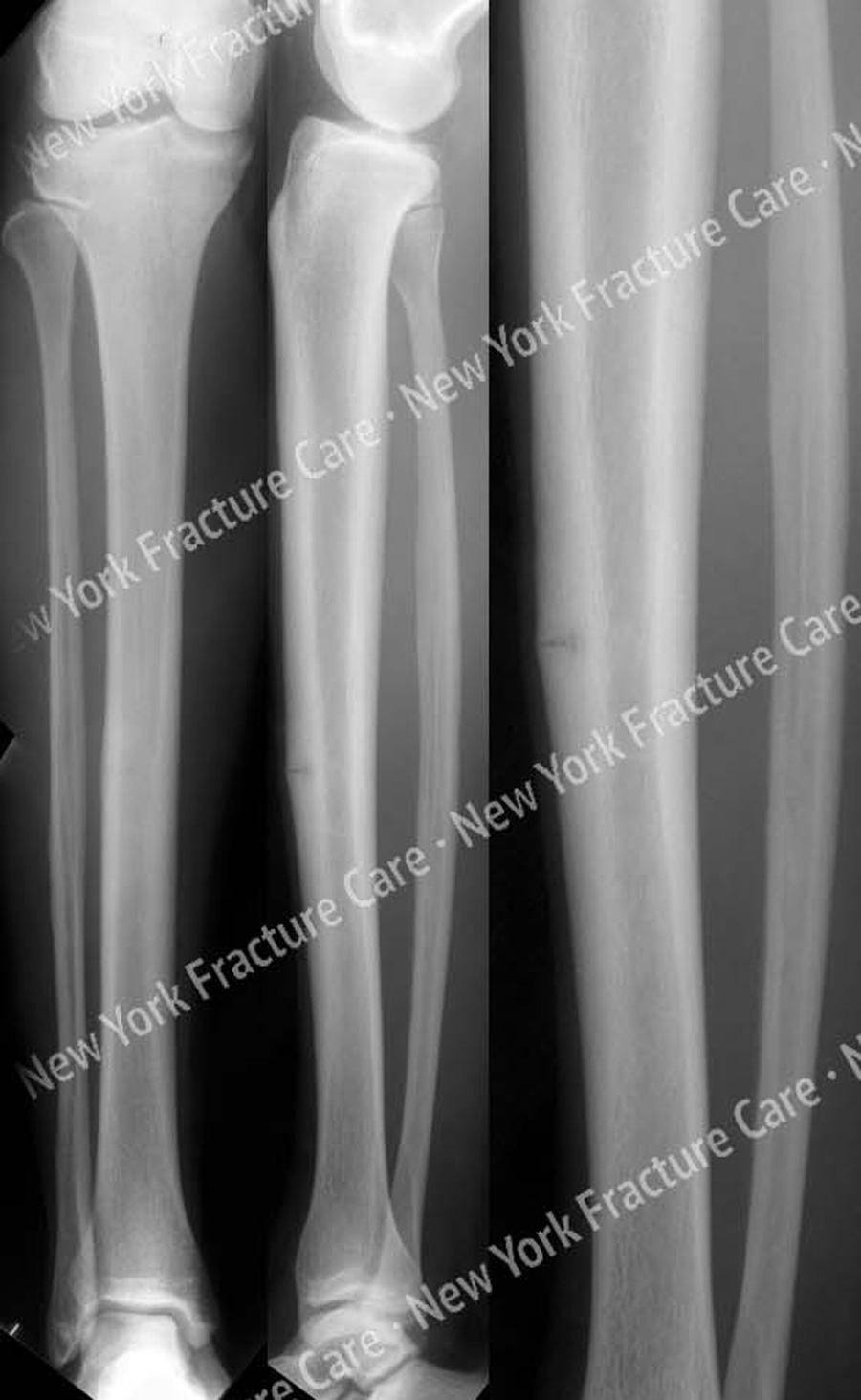
Radiographs revealed a radiolucent line involving the anterior cortex of her tibia at the mid-shaft level. She was treated with anterior tension band plating and bone graft using a locking plate.
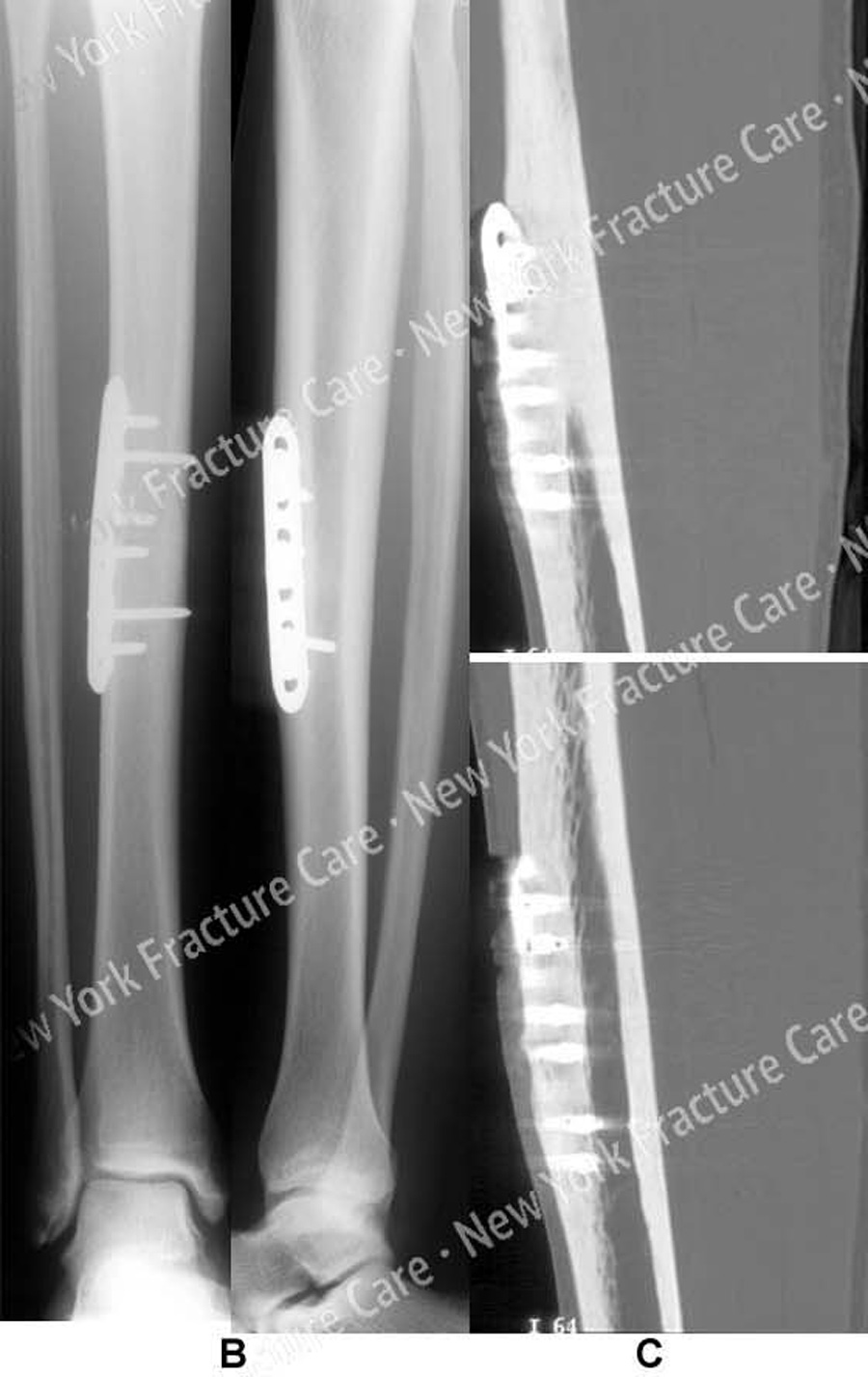
At 10 weeks follow up her radiographs illustrated healing of the stress fracture and she resumed full activities. At 4 months postoperatively she had resumed training for competition. At 1 year follow-up she was completely asymptomatic.
-
Figure A
Anteroposterior (AP) and lateral radiographs reveal a stress fracture of the anterior tibial cortex (arrows). -
Figure B, C
(B) AP and lateral radiographs 8 months following surgery revealing a healed tibia stress fracture.
(C) CT scan images at 8 months confirm fracture healing.
Tags: Stress Fracture, Tibia Fracture


