Tibia fractures (lower extremity)
A 15-year-old female, avid soccer and volleyball player, noted progressive onset of right-sided shin pain during her athletic training over the course of several weeks.
Radiographs taken at a local hospital revealed a minimally displaced mid-shaft tibia stress fracture. She was initially treated with cast immobilization for 3 months. She was referred to us at 6 months for treatment of a malunion with bridging callus on the lateral cortex and 13° of valgus angulation.
Operative treatment was planned and performed including correction of the deformity and insertion of an intramedullary(IM) nail and locking screws.
She returned for regular follow-up visits and at 3 months following surgery she had excellent radiographic and clinical results including a healed tibia fracture, resolution of pain and full range of motion of the knee and ankle joints. She returned to all pre-injury activities at 4 months. At 1 year following surgery the hardware was removed.
-
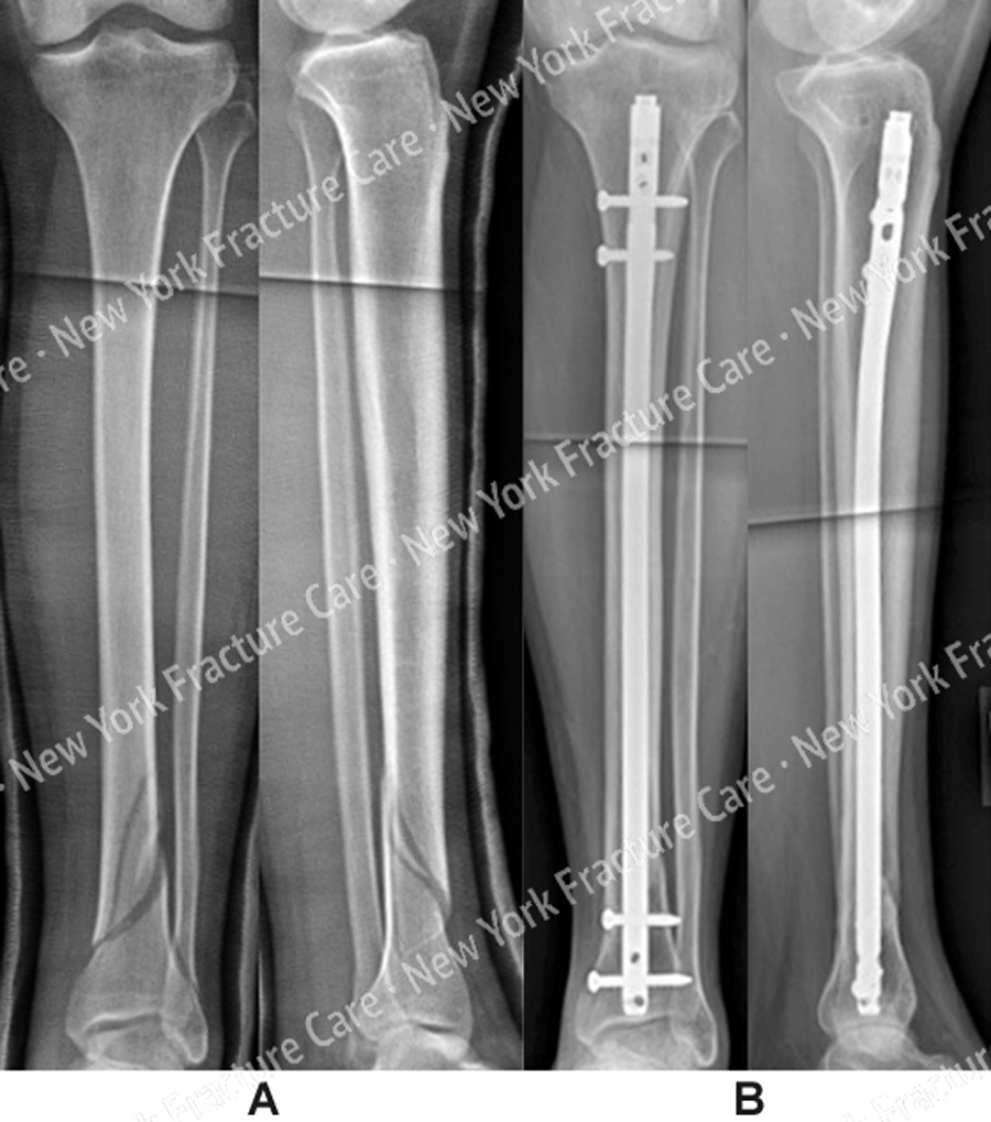
Figure A, B
(A) Anteroposterior (AP) and lateral radiographs reveal a minimally displaced mid-shaft tibia stress fracture (left); AP and lateral radiographs at 6 months following the injury illustrating a malunion with 13° of valgus deformity (right).
(B) AP and lateral radiographs 6 months following surgery illustrating a healed tibial malunion in excellent alignment (left); Intraoperative fluoroscopic images following removal of hardware at 12 months (right).
Tags: Stress Fracture, Tibia Fracture

