Fractures with osteoporosis
A 48-year-old female was referred to Dr. David L. Helfet 2 days following a fall from a standing height onto her left upper extremity.
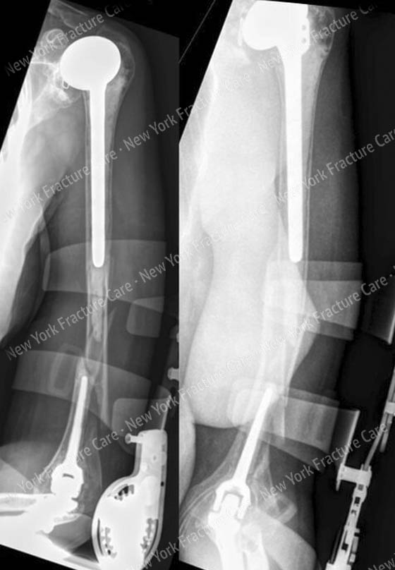
Radiographs revealed a periprosthetic fracture above a cemented total elbow replacement and below a cemented humeral component of a total shoulder replacement. Her history was also significant for osteoporosis.
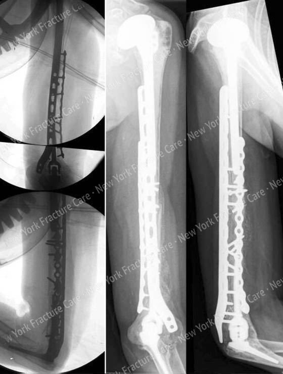
Open reduction and internal fixation (ORIF) was performed with placement of a 3.5mm extra-articular distal humeral locking plate posterior-laterally and a 3.5mm reconstruction plate along the medial column in a 90-90 construct with multiple interfragmentary lag screws.
She returned for routine follow-up and at 7 months radiographs illustrate a healed periprosthetic humerus fracture and she has returned to activities of daily living with resolution of pain.
-
Figure A
Anteroposterior (AP), oblique and lateral radiographs revealing a periprosthetic humerus fracture between a cemented long stemmed humeral component and a cemented standard length elbow component. -
Figure B
Anteroposterior and lateral fluoroscopic images demonstrating adequate fixation and alignment following open reduction and internal fixation. Radiographs at 7 months postoperatively demonstrate a healed periprosthetic humerus fracture.
*Carroll EA, Lorich DG, Helfet DL: Surgical management of a periprosthetic fracture between a total elbow and total shoulder prostheses: a case report. J Shoulder Elbow Surg. 2009 18(3):e9-12.
Tags: Broken Shoulder, David Helfet MD, Elbow Fracture, Osteoporosis


